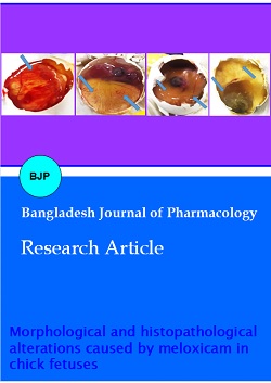
Morphological and histopathological alterations caused by meloxicam in chick fetuses
DOI:
https://doi.org/10.3329/bjp.v17i4.62233Keywords:
Chick embryo, Meloxicam, PregnancyAbstract
During pregnancy, the administration of certain drugs can cause harm to the unborn child. The purpose of the present study was to investigate the impact of meloxicam on morphological and histopathological changes in chick embryo. Forty-eight eggs were divided into four groups. After 12 days of incubation (37.8ºC, humidity - 65%), the groups were given meloxicam at varying doses. Fetuses were then removed after 48 hours. The results show that administration of high doses of meloxicam produced weakly or absence of vitelline vascularization. Also, meloxicam application instigated fetal deformities such as growth retardation, subcutaneous hemorrhage, very thin skin and exencephaly. Many histopathological alternations were noted in the retina, liver and kidney tissues of chick fetus of treated groups with meloxicam compared to the control group. The high doses of meloxicam caused many morphological and histopathological changes in the chick fetus. The safety of meloxicam was not established in this study.
Downloads
228
139
References
AbdRabou MA, Al-Ghamdi FA, Al-Otaibi AM , Gewily DI. Histopathological, histochemical and immunological studies on fetal pancreatic tissue of rats treated with carisoprodol. Int J Pharmacol. 2021; 17: 506-16.
AbdRabou MA. Effect of different doses of meloxicam on the development of chick embryo. Int J Pharmacol. 2021; 17: 474-81.
Al-Rekabi F, Abbas D, Hadi N. Effects of subchronic exposure to meloxicam on some hematological, biochemical and liver histopathological parameters in rats. Iraqi J Vet Sci. 2009; 23: 249-54.
Ami N, Bernstein M, Boucher F, Rieder M, Parker L. Folate and neural tube defects: The role of supplements and food fortification. Paediatr Child Health. 2016; 21: 145-49.
Antonucci R, Zaffanello M, Puxeddu E, Porcella A, Cuzzolin L, Pilloni MD, Fanos V. Use of non-steroidal anti-inflammatory drugs in pregnancy: Impact on the fetus and newborn. Curr Drug Metab. 2012: 13: 474-90.
Burdan F. Comparison of developmental toxicity of selective and non-selective cyclooxygenase-2 inhibitors in CRL: (WI)WUBR Wistar rats-DFU and piroxicam study. Toxicology 2005; 211: 12-25.
Burukoglu D, Baycu C, Taplamacioglu F, Sahin E, Bektur E. Effects of nonsteroidal anti-inflammatory meloxicam on stomach, kidney, and liver of rats. Toxicol Ind Health. 2016; 32: 980-86.
Cetinkal A, Colak A, Topuz K, Demircan MN, Simsek H. The effects of meloxicam on neural tube development in the early stage of chick embryos. Turk Neurosurg. 2010; 20: 111-16.
Dzięcioł M, Szpaczek A, Uchańska O, Niżański W. Influence of a single dose of meloxicam administrated during canine estrus on progesterone concentration and fertility: A clinical case study. Animals 2022; 12: 655.
Elkomy AA, Salem MA, Kandeil AM, Hassan A, Elhemiely AA. Teratogenic effect of meloxicam on pregnant rats: Implication of organogenesis period. Benha Vet Med J. 2018; 35: 317-27.
Erdem H , Guzeloglu A. Effect of meloxicam treatment during early pregnancy in Holstein heifers. Reprod Domestic Anim. 2010; 45: 625-28.
Geliflimine EN. The effects of meloxicam on neural tube development in the early stage of chick fetuses. Tur Neurosurg. 2010; 20: 111-16.
Karkoszka M, Rok J, Banach K, Kowalska J, Rzepka Z, Wrz-eśniok D. The assessment of meloxicam phototoxicity in human normal skin cells: In vitro studies on dermal fibroblasts and epidermal melanocytes. Molecules 2022: 27: 4215.
Mahaprabhu R, Bhandarkar AG, Jangir BL. Ameliorative effect of Ocimum sanctum on meloxicam induced toxicity in Wistar rats. Toxicol Int. 2011; 18: 130-36.
Ofori B, Oraichi D, Blais L, Rey E, Bérard A. Risk of congenital anomalies in pregnant users of non-steroidal anti-inflammatory drugs: A nested case-control study. Birth Defects Res. Part B: Dev Reprod Toxicol. 2006; 77: 268-79.
Oliveira ÉD., Azevedo RD, Bonfilio R, de Oliveira DB, Ribeiro GP, de Araújo MB. Dissolution test optimization for meloxicam in the tablet pharmaceutical form. Braz J Pharm Sci. 2009; 45: 67-73.
Sachdeva P, Patel BG, Patel BK. Drug use in pregnancy, a point to ponder. Indian J Pharm Sci. 2009; 71: 1-7.
Stephansson O, Granath F, Haglund A. Ekbom A, Kieler H. Drug use during pregnancy in Sweden-assessed by the prescribed drug register and the medical birth register. Clin Epidemiol. 2011; 3: 43-50.
Sturmer T, Elseviers MM, De Broe ME. Non-steroidal anti-inflammatory drugs and the kidney. Curr Opin Nephrol Hypertens. 2001; 10: 161-63.
Sulaiman AH, Jarjees MT. The effect of diclofenac and meloxicam on liver and kidney of male rats. A histomorphometrical study (Doctoral dissertation, PhD Thesis]. 2010. Collage of Medicine, University of Mosul).
Sun AJ, Li S, Zhang CA, Jensen TK, Lindahl-Jacobsen R, Eisen-berg ML. Parental comorbidity and medication use in the USA: A panel study of 785 000 live births. Hum Reprod. 2020; 35: 669-75.
Temiz C, Temiz P, Demirel A, Sayın M, Umur AŞ, Özer FD. Effect of sodium phenytoin concentration on neural tube development in the early stages of chicken embryo development. J Clin Neurosci. 2009; 16: 307-11.
Urban M . Cox-2 specific inhibitors offer improved advantages over traditional NSAIDs. Orthopedis 2000; 23: 61-64.

Downloads
Published
How to Cite
Issue
Section
License
Copyright (c) 2022 Mervat AbdRabou, Aljohara Al- Otaibi

This work is licensed under a Creative Commons Attribution 4.0 International License.
Authors who publish with this journal agree to the following terms:
- Authors retain copyright and grant the journal right of first publication with the work simultaneously licensed under a Creative Commons Attribution License that allows others to share the work with an acknowledgement of the work's authorship and initial publication in this journal.
- Authors are able to enter into separate, additional contractual arrangements for the non-exclusive distribution of the journal's published version of the work (e.g., post it to an institutional repository or publish it in a book), with an acknowledgement of its initial publication in this journal.
- Authors are permitted and encouraged to post their work online (e.g., in institutional repositories or on their website) prior to and during the submission process, as it can lead to productive exchanges, as well as earlier and greater citation of published work (See The Effect of Open Access).
