Barraquer–Simons syndrome: A case report

Authors

DOI:

Keywords

lipodystrophies, Barraquer–Simons syndrome, fat wasting

Correspondence

Mita Dutta 
Email: mitadutta42@gmail.com

Publication history

Received: 22 Dec 2024
Accepted: 30 Mar 2025
Published online: 20 Apr 2025

Handling editor

Reviewers

Funding

None

Ethical approval

Ethical approval was not sought because this is a case report. However, written informed consent was obtained from the patient for publication of this case report and any accompanying images. 

Trial registration number

Not applicable

Copyright

© The Author(s) 2025; all rights reserved
Published by Bangabandhu Sheikh
Mujib Medical University

Introduction
Barraquer-Simon syndrome (BSS) is an acquired partial lipodystrophy characterised by bilateral symmetrical loss of fat from the upper extremities [1]. Although mutations in the lamin B2 gene have been reported, they are not definitive for diagnosis [2]. These patients exhibit less pronounced features of insulin resistance compared to other lipodystrophies [3]. Diagnosis of this rare condition can be done by the presence of the essential component of the disease along with other supporting evidence [4].
Key messages
Barraquer–Simons syndrome is a rare condition characterised by the loss of upper body fat and hypertrophy of lower body fat, along with pronounced psychological stress due to facial fat loss but less pronounced features of insulin resistance and its complications. Pioglitazone and cosmetic surgery are the primary treatment options.
Case description and management
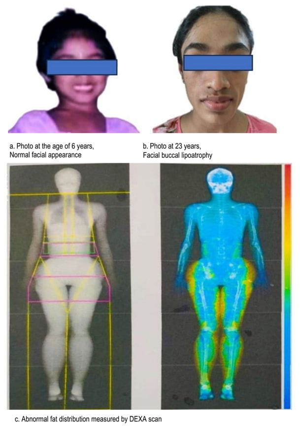
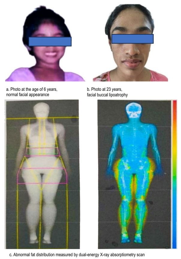
A 23-year-old Bangladeshi female, the second offspring of healthy, non-related parents, was referred to the endocrinology department for facial fat loss. She had oligomenorrhea since the onset of her menarche. At puberty, she noticed a gradual decline in her facial adipose tissue. Apart from viral illness in her teen years, she had no significant family or drug history. On physical examination, she had large zygomatic arches and disappearance of buccal fat pads (Figure 1). She had mild hirsutism, mild acanthosis nigricans, and umbilical hernia. Her waist and hip circumference were 69 and 90 cm, respectively. There were no features of autoimmune disorders, clitoromegaly or hepato-splenomegaly. Her routine blood, urine tests, and hormonal profiles were within normal limits. Her antinuclear antibody was also negative. She had dyslipidemia in the form of low high-density lipoprotein cholesterol (40 mg/dL), high low-density lipoprotein cholesterol (230 mg/dL), and a marginally elevated triglyceride level (351 mg/dL). Her complement C3 level was low at 0.767 g/L (reference ranges 0.9-1.8 g/L). Although her glycemic profiles were within normal limits, she had elevated fasting insulin level of 35.36 micro U/mL (reference values: 3.21-16.32 micro U/mL) and an elevated homeostatic model assessment for insulin resistance of 6.37 (reference ranges: <1.9). Fat mass was measured using a dual-energy X-ray absorptiometry scan. She was treated with pioglitazone to improve the distribution of fat in her body. Rosuvastatin was started to treat her dyslipidemia. She was also counseled about diet, exercise, and lifestyle changes.
 
As this young woman was facing distress due to her facial appearance, we referred her to a plastic surgeon for facial fat grafting to enhance her appearance. We kept her on regular follow-up to monitor her metabolic and renal parameters as there is an increased chance of development of chronic kidney disease due to membranoproliferative glomerulonephritis. 

Categories

Number (%)

Sex


   Male

36 (60.0)

   Female

24 (40.0)

Age in yearsa

8.8 (4.2)

   Education


   Pre-school

20 (33.3)

   Elementary school

24 (40.0)

   Junior high school

16 (26.7)

Cancer diagnoses


   Acute lymphoblastic leukemia

33 (55)

   Retinoblastoma

5 (8.3)

   Acute myeloid leukemia

4 (6.7)

   Non-Hodgkins lymphoma

4 (6.7)

   Osteosarcoma

3 (5)

   Hepatoblastoma

2 (3.3)

   Lymphoma

2 (3.3)

   Neuroblastoma

2 (3.3)

   Medulloblastoma

1 (1.7)

   Neurofibroma

1 (1.7)

   Ovarian tumour

1 (1.7)

   Pancreatic cancer

1 (1.7)

   Rhabdomyosarcoma

1 (1.7)

aMean (standard deviation)

Categories

Number (%)

Sex


   Male

36 (60.0)

   Female

24 (40.0)

Age in yearsa

8.8 (4.2)

Education


   Pre-school

20 (33.3)

   Elementary school

24 (40.0)

   Junior high school

16 (26.7)

Cancer diagnoses


Acute lymphoblastic leukemia

33 (55)

Retinoblastoma

5 (8.3)

Acute myeloid leukemia

4 (6.7)

Non-Hodgkins lymphoma

4 (6.7)

Osteosarcoma

3 (5)

Hepatoblastoma

2 (3.3)

Lymphoma

2 (3.3)

Neuroblastoma

2 (3.3)

Medulloblastoma

1 (1.7)

Neurofibroma

1 (1.7)

Ovarian tumour

1 (1.7)

Pancreatic cancer

1 (1.7)

Rhabdomyosarcoma

1 (1.7)

aMean (standard deviation)

Pain level

Number (%)

P

Pre

Post 1

Post 2

Mean (SD)a pain score

4.7 (1.9)

2.7 (1.6)

0.8 (1.1)

<0.001

Pain categories


   No pain (0)

-

(1.7)

31 (51.7)

<0.001

   Mild pain (1-3)

15 (25.0)

43 (70.0)

27 (45.0)


   Moderete pain (4-6)

37 (61.7)

15 (25.0)

2 (3.3)


   Severe pain (7-10)

8 (13.3)

2 (3.3)

-


aPain scores according to the visual analogue scale ranging from 0 to 10; SD indicates standard deviation

Figure 1 Characteristic features of Barraquer–Simons syndrome. 
Yellow color represents increased fat mass in lower body.
Discussion
BSS is rarely observed in Asian countries, with only 250 cases reported in the literature [35, 6] To our knowledge, no comparable cases have been recorded from Bangladesh thus far.

BSS has a female preponderance, with a median age of presentation at 25 years [4]. This disease is believed to have originated from unknown causes, but it exhibits implications of viral illnesses, such as chickenpox and measles, as well as autoimmune associations with systemic lupus erythematosus (SLE), vasculitis, and other conditions. Although 80% of cases are associated with autoimmune diseases, most cases can develop them many years after the initial diagnosis of lipodystrophy (systemic lupus erythematosus, 4–37 years; vasculitis, 17–28 years) [4]. BSS is atypical from other lipodystrophies. Although the predominant abnormality is hypertriglyceridemia, hypercholesterolemia can be present in a minority of cases [4].
 
The diagnosis of BSS was made using the recognised standards of Mishra et al., in which the hallmark feature is the loss of fat from the face and chest, accompanied by the deposition of excess fat in the hips, thighs, and legs. These physical findings can be confirmed by the imaging results of a dual-energy X-ray absorptiometry scan, which is considered the gold standard for measuring body fat [4]. Evidence of insulin resistance and hypocomplementemia enhances further confirmation. Most authors suggested a finding of low complement in the cases of BSS [4, 5].  
 
In cases of BSS, patients mainly raise concern about their facial appearance. This finding is absent in cases of facial puffiness due to chronic kidney disease as there is an association with membranoproliferative glomerulonephritis [4]. However, only 22% of cases developed it after a median of 8 years following the onset of lipodystrophy [4]. 
 
Pioglitazone has particular importance in cases of acquired partial lipodystrophy as it improves fat distribution and is associated with a reversal of chronic inflammation and insulin resistance [5]. Cosmetic restoration through autologous fat grafting is a promising treatment option, with sustained positive outcomes observed in follow up among patients with BSS [6].
BSS is a rare condition that has profound psychological and clinical effects; therefore, the authors emphasise the importance of detecting and evaluating patients with acquired partial lipodystrophy.
 
Acknowledgements
None
Author contributions
Manuscript drafting and critical revision: MD, MAS, MAI, HB. Approval of the final version of the manuscript: MD, MAS, MAI, HB. Guarantor of accuracy and integrity of the work: MD, MAS, MAI, HB.
Conflict of interest
We do not have any conflict of interest.
Data availability statement
We confirm that the data supporting the findings of the study will be shared upon reasonable request. 
Supplementary file
None
    References
    1. Brown RJ, Araujo-Vilar D, Cheung PT, Dunger D, Garg A, Jack M, Mungai L, Oral EA, Patni N, Rother KI, Von Schnurbein J. The diagnosis and management of lipodystrophy syndromes: a multi-society practice guideline. The Journal of Clinical Endocrinology & Metabolism. 2016 Dec 1;101(12):4500-4511. doi: https://doi.org/10.1210/jc.2016-2466
    [PubMed]     [Google Scholar]
    2. Simsek‐Kiper PO, Roach E, Utine GE, Boduroglu K. Barraquer–Simons syndrome: A rare clinical entity. American Journal of Medical Genetics Part A. 2014 Jul;164(7):1756-1760. doi: https://doi.org/10.1002/ajmg.a.36491
    [PubMed]     [Google Scholar]
    3. Oliveira J, Freitas P, Lau E, Carvalho D. Barraquer–Simons syndrome: a rare form of acquired lipodystrophy. BMC research notes. 2016 Dec; 9:1-4. doi: https://doi.org/10.1186/s13104-016-1975-9
    [PubMed]     [Google Scholar]
    4. Misra A, Peethambaram A, Garg A. Clinical features and metabolic and autoimmune derangements in acquired partial lipodystrophy: report of 35 cases and review of the literature. Medicine. 2004 Jan 1;83(1):18-34. doi: https://doi.org/10.1097/01.md.0000111061.69212.59
    [PubMed]     [Google Scholar]
    5. Corvillo F, Ceccarini G, Nozal P, Magno S, Pelosini C, Garrido S, López-Lera A, Moraru M, Vilches C, Fornaciari S, Gabbriellini S, Santini F, Araújo-Vilar D, López-Trascasa M. Immunological features of patients affected by Barraquer-Simons syndrome. Orphanet J Rare Dis. 2020 Jan 10;15(1):9. doi: https://doi.org/10.1186/s13023-019-1292-1 
    [PubMed]     [Google Scholar]
    6. Kumar R, Aneja SS, Seth A, Taluja V. Partial lipodystrophy in a boy. Indian Pediatrics. 2000 Jan 1;37(1):93-96. URL: https://www.indianpediatrics.net/jan2000/case3.htm
    [PubMed]     [Google Scholar]